DENTISTRY and OROFACIAL SURGERY
of EXOTIC COMPANION MAMMALS
- ONLINE COURSE -


Sponsored by
Rollover/click or Tap
over the tab
for more info
and the preview
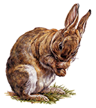
RABBIT
DIAGNOSTIC IMAGING
This session of the course is dedicated entirely to diagnostic imaging modalities for rabbit dentistry.
Standard imaging (radiography, oral endoscopy), and advanced imaging (computed tomography, magnetic resonance) will be extensively discussed in regard of both normal aspects and pathologic findings.
Radiography of the Head and Teeth (93 min.)

This extensive lecture is entirely focused on radiography for diagnosis of rabbit dental disease.
It includes:
- the radiographic equipment;
- the patient positioning;
- the extraoral projections including additional projection and the normal radiographic anatomy;
- the use of diagnostic lines;
- the intraoral views;
- an overview of radiographic abnormalities
This is just a short preview!
Register to view the full lecture
Inspection of the Oral Cavity and Oral Endoscopy (31 min.)
This is just a short preview!
Register to view the full lecture
This lecture focuses on the inspection of the oral cavity under anesthesia, with the aid of rigid endoscopy (stomatoscopy).
Subtopics will be discussed as follows:
- instruments for inspection of the oral cavity, including the specific table top mouth gag/restrainer;
- equipment for rigid endoscopy;
- stomatoscopy of the normal patient;
- an overview of dental and oral abnormalities diagnosed via stomatoscopy
Computed Tomography (CT) (78 min.)
This lecture is entirely focused on CT as advanced imaging modality for diagnosis of dental disease in rabbits.
It includes:
- the basic principles of CT;
- different types of CT scan units;
- patient positioning;
- the scanning planes and the slice thickness;
- the DICOM viewers;
- an overview of dental, oral and facial abnormalities diagnosed via CT scan
This is just a short preview!
Register to view the full lecture
Practical Session: Viewing CT Scan (90 min.)
This is just a short preview!
Register to view the full lecture
This session shows in detail the use of DICOM software for viewing CT scans, including basic functions such as 2D orthogonal MPR, 3D curved MPR, 3D surface and volume rendering. Clinical cases will be displayed and discussed
Magnetic Resonance Imaging (MRI) (31 min.)
his lecture is entirely focused on MRI as advanced imaging modality for diagnosis of complications of dental disease in rabbits.
It includes:
- the basic principles of MRI;
- the standard sequences;
- an overview of dental, oral and facial abnormalities diagnosed via MRI scan;
- comparison with CT scan
This is just a short preview!
Register to view the full lecture
The format of the Dentistry and Orofacial Surgery of Exotic Companion Mammals course is a copyright of Vittorio Capello, DVM © 2018